K.J.Leonard & Suggs (1974)
What is Exserohilum?
The genus Exserohilum belongs to the family Pleosporaceae of the phylum Ascomycota. They are filamentous fungi, classified into the group of dematiaceous species (darkly pigmented) [1, 2]. Exserohilum genus includes approximately thirty-five mostly asexually reproducing fungi, of which sexually reproducing species are assigned to the genus Setosphaeria [3].

Exserohilum species are saprophytic fungi, commonly found in soil and grass, occurring mainly in warm tropical and subtropical regions. Some species are human pathogens, causing a wide range of localized and systemic infections in both immunosuppressed and immunocompetent patients [1].
Cultural and microscopic features of Exserohilum species
Exserohilum species grow rapidly on potato dextrose agar (PDA), producing woolly colonies. Colonies are initially pale, but quickly turn dark gray to black with a black reverse [3].
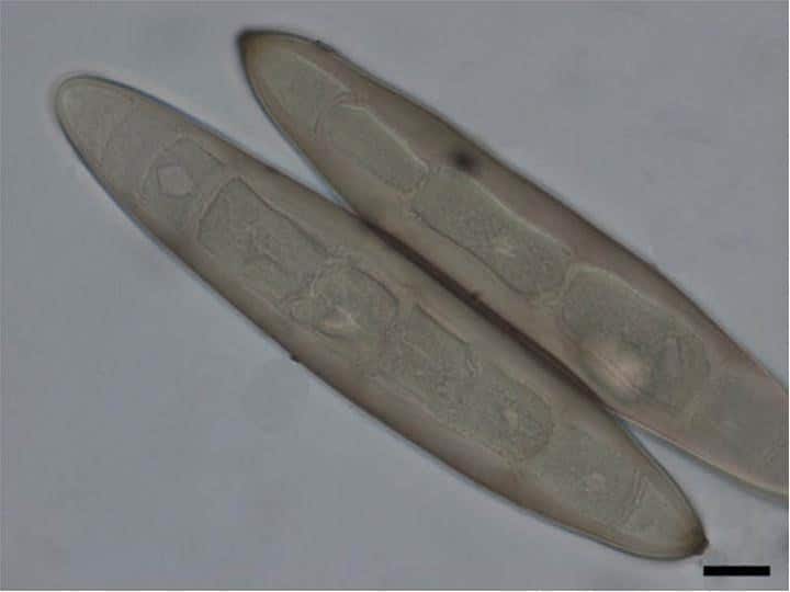
Microscopically, Exserohilum species are characterized by septate pigmented hyphae. Conidiophores are septate, non-branched, bending at a sharp angle and becoming paler near the tip. Conidia differ in size between strains and are colored in various shades of brown (Fig. 1) [2, 3]. They may be straight or curved, divided into 4–14 septae [2]. The most distinguishing characteristic of the genus is the protruding hilum, a scar left on a conidium after detachment [2, 3]. Above the hilum is usually a thickened, dark septum [4].
How many Exserohilum species exist?
Among approximately thirty-five species, the Exserophilum genus comprises three human pathogens: E. rostratum, E. longirostratum, and E. mcginnisii [2]. E. rostratum is reported to cause about 60% of Exserohilum infections, which are manifested as systemic (73%), dermal (25%), corneal (16.7%), and hypodermal (10.4%) disease. Recent molecular studies have shown a high similarity between these three taxa, suggesting that they could be the same species [1].
What are Exserohilum human infections?
According to the reports [1], the most common kind of infection caused by Exserohilum spp. is systemic, followed by dermal (cutaneous) and corneal infections. Systemic infection primarily affects the sinuses and lungs due to inhalation of the pathogen. Skin and eye infections are most commonly caused by wounds from contaminated plants (wound infections) [1]. The majority of reported patients were from the United States, while others were from India, Israel, East Asia, and Canada [1]. Most of them had an underlying condition or were immunosuppressed [1].
The tragic outbreak of E. rostratum meningitis happened in 2012 in the United States, caused by contaminated steroid injections [1, 5]. Contaminated injection solutions exposed more than 13,000 people to infection, with 741 cases of confirmed infection and 55 cases of fatal outcomes [5]. Besides meningitis, joint infections were also reported [1]. Before this outbreak, the only case of meningitis caused by Exserohilum had been published [1].
What are symptoms and treatment of Exserohilum infections?
Symptoms of infections caused by Exserohilum species are similar to those caused by other dematiaceous fungi. These infections have a common name – phaeohyphomycosis [7]. Symptoms of systemic infections include invasive sinusitis, a lung disease with fungal masses, osteomyelitis, mycotic arthritis, bone necrosis, endocarditis, and disseminated infection [7]. Eye infection is manifested as keratitis with a darkly pigmented lesion and feathery edges, sometimes with corneal ulcers [8]. Skin infection begins with localized lesions, which can be followed by subcutaneous nodules or abscesses [7].
According to published data [1], treatment with amphotericin B alone, or combined with an azole, has shown the best results in treating Exserohilum infections.
How to prevent Exserohilum infections?
Unfortunately, there is no prevention and control suggested when it comes to infection caused by Exserohilum species and other dematiaceous fungi. Considering their cosmopolitan and ubiquitous nature, it is not feasible to eradicate them from the environment [8].
However, people should be careful when handling plants and soil to avoid trauma from contaminated material. Also, very strict quality control procedures must be implemented in the drug production industry to avoid infection outbreaks from happening again.
Exserohilum as plant pathogens
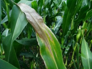
Exserohilum turcicum causes the Turcicum leaf blight (TLB), also known as Northern corn leaf blight (NCLB). TLB is a foliar corn disease, causing significant yield losses in susceptible maize hybrids. This disease causes the characteristic cigar-shaped or elliptical necrotic grayish-green lesions on corn leaves, spanning up to seven inches long (Fig. 2). As the infection progresses, the lesions grow together and form larger areas of dead tissue. If left untreated, the lesions may spread so much that the foliage is eventually destroyed. If severe infections are present 2–3 weeks after silking in field corn, the grain yield can be reduced by as much as 30% [9,10].
.jpg)
Did you know?
The #1 toxic mold type found in homes is the Penicillium/Aspergillus mold group?! Find out more exciting mold stats and facts inside our mold statistics page.
References
- Katrahgkou, A. et al. (2014). Exserohilum infections: Review of 48 cases before the 2012 United States outbreak. Medical Mycology. 52: 376–386
- Sutton, D. A., Rinaldi, M. G., & Sanche, S. E. (2009). Dematiaceous fungi. In Clinical Mycology (pp. 329-354). Elsevier Inc.
- Exserohilum Retreated from: www.drfungus.org
- Exserohilum. Retreated from: adelaide.edu.au
- Ritter, J. M. et al. (2013). ExserohilumInfections Associated with Contaminated Steroid Injections. A Clinicopathologic Review of 40 Cases. The American Journal of Pathology. 183(3): 881–892.
- Chowdhary, A. et al. (2014). ESCMID and ECMM joint clinical guidelines for the diagnosis and management of systemic phaeohyphomycosis: diseases caused by black fungi
- Chaindaroon, W. et al. (2019). Exserohilum rostratum Keratitis in a Patient with Human Immunodeficiency Virus. Case Reports in Ophthalmology 10(1):127-133
- Matsumoto, T., C. R. Cooper, Jr. and J. Szaniszlo (2011). Chromoblastomycosis and Phaeohyphomycosis. In: R. L. Guerrant, D. H. Walker and P. F. Weller (ed.) Infectious Diseases: Principles, Pathogens, and Practice, Third Edition. Saunders Elsevier, Oxford, U.K pp. 569-572.
- Welz, H. G., and H. H. Geiger. “Genes for resistance to northern corn leaf blight in diverse maize populations.” Plant breeding 119.1 (2000): 1-14.
- Wise, Kiersten (2011). “Diseases of Corn: Northern Corn Leaf Blight”. Purdue University Extension Publication. Purdue University.
extension.purdue.edu
Get Special Gift: Industry-Standard Mold Removal Guidelines
Download the industry-standard guidelines that Mold Busters use in their own mold removal services, including news, tips and special offers:

Written by:
Jelena Somborski
Mycologist
Mold Busters
Edited by:
Dusan Sadikovic
Mycologist – MSc, PhD
Mold Busters